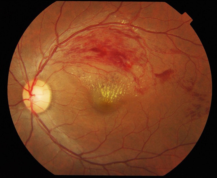
Central vein occlusion of the retina is a disease that appears when there is an obstruction in the flow of deoxygenated blood from the retina. The authors of this work have studied the efficacy of a new surgical technique against this disease in individuals with different ages to observe the different efficacy of the treatment depending on the patient’s age.
This article analyzes the efficacy of a recent surgical technique (radial optic neurotomy or RON) in the treatment of central vein occlusion of the retina (OVCR) in 2 groups of patients: group 1 <of 50 years of age I group 2> 50.
OVCR is the second retinal vascular pathology after diabetic retinopathy and occurs around 60 years of age on average. It is produced when there is an obstruction in the return of deoxygenated blood from the retina, which causes the accumulation of fluid (edema) in the area of maximum vision of the retina (macula), as well as areas of infarction. The main risk factors are high blood pressure, diabetes mellitus and chronic glaucoma.
When it occurs in younger patients, it is considered a more benign disease and that in theory is caused by some underlying disease. Often the evolution of the picture is different although there are no conclusive publications on the prevalence of the majority of hereditary risk factors.
The natural history of the OVCR has shown that the evolution of this disease depends mainly on the level of vision that patients present at the time of diagnosis so that those patients who have a vision below 0.1-0.2 have a visual prognosis very poor.
In the study presented by the authors, 43 patients have an initial vision <0.2; risk factors are analyzed and all patients undergo this surgical intervention that aims to improve the circulation of the central vein of the retina. Visual evolution is monitored for 12 months after treatment and decreased retinal edema (using the OCT, a retinal scanner that measures thickness).
There are two types of retinal venous occlusion:
Retinal vein occlusion (OVCR): when the main vein of the eye is blocked (located in the optic nerve)
Venous branch occlusion of the retina (ORVR): when one of the smallest branches of the vessels connected to the main vein becomes blocked
Central vein occlusion of the retina (OVCR): The main vein of the eye is clogged and this causes bleeding (bleeding) in the retina.
Venous branch occlusion of the retina (ORVR): A smaller branch of vessels connected to the main vein is clogged and this causes bleeding in parts of the retina
Certain diseases increase the risk of retinal venous occlusion, such as:
- the diabetes
- glaucoma
- high blood pressure
- an age-related vascular disease (blood vessel)
- blood disorders
Among the results obtained the fact that vascular risk factors were more frequent in group 2 (as was already known) and that among the patients in group 1, the number of hereditary risk factors was low.
In relation to the visual results, these were better in the youth group; This hehco occurs in other pathologies of the retina since in young patients the retina has a greater capacity to recover its normal function. In both groups, retinal oedema and haemorrhages improved. Despite the best evolution in the youth group, the pathology cannot be considered “benign”, since the final vision is equally poor.
Therefore, the study concludes that in both groups of patients the technique is effective, but with better functional results in young people.
